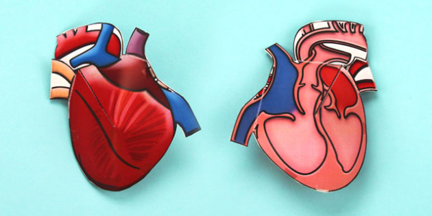
3D models within Anatomy & Physiology Revealed (APR) 4.0 allow students to explore the spatial relationships between structures by using a mouse or keyboard navigation. The heart 3D model in APR 4.0 is accessed by clicking the below icon in Module 15: Regional Anatomy, Module 7: Nervous, or Module 9: Cardiovascular.
- A bioprinted embryonic human heart model at 84 times the actual size. This is one of two different kinds of models Vahid Serpooshan is developing that are 3D printed using soft, flexible hydrogel materials infused with cells from specific patients.
- The Zygote Solid 3D Heart Model is the most comprehensive and medically accurate 3D CAD model of the human heart. It is a highly accurate model created for use within all major engineering applications for finite element analysis, product design, and visualization. This carefully crafted model is based on human CT and MRI scans, and accurately.
- Mar 20, 2018 Vertices: 97.1k. More model information. Anatomical cross section of the human heart showing the internal working and complexity of one of the hardest working organs in the body. Published 3 years ago. Art & abstract 3D Models.
If not yet familiar with how to navigate and interact with the 3D models in APR, please click on the help icon (indicated by yellow arrow) in the lower left corner, which will launch a tutorial explaining the functionality of the 3D model viewer.
Human Heart 3D models for download, files in 3ds, max, c4d, maya, blend, obj, fbx with low poly, animated, rigged, game, and VR options. Our anatomical heart models feature a unique and interactive hands-on design.Each of our human heart models offer a 3D heart depiction, which allows doctors, students, and patients the opportunity to explore various pathways and system functions of the organ itself, diseases of the heart, and surgical procedures.
The “Create link” icon is a quick way to create a customized bookmark to allow someone else to access the same exact location in APR 4.0.
How do I use the “Create link” icon?
Once you have the location you’d like to share, click the clipboard icon (blue arrow) to copy the link to your clipboard. Dps velocity editing software free download. Paste this link into a document, presentation slide, website, or in your learning management system (LMS). Someone trying to access this link simply needs to first log into APR (since it is a password-protected website) or already have a browser tab open to APR, then click on the link to be taken to this same exact location in APR. This “Create link” feature works with the 3D model and in any other mode in APR such as dissection or histology.
Below are links to different views in the heart 3D model that may benefit your students as they learn and study heart anatomy.
Anterior view of heart focused on coronary vessels
This view allows a student to observe the major coronary vessels of the heart. The external anatomy of the heart has been set to “Transparent” but could easily be changed to “Show”. With the 3D model, a student can tilt and rotate the heart to get an optimal view of each coronary vessel. The transparent setting of the heart 3D model makes it easier to see the branching of different vessels.
https://anatomy.mheducation.com/html/apr.html?animal=human&mod=4178
Anterior view of heart focused on external heart anatomy
By changing the visibility of the menu “External surface of heart” from Transparent to Show, the external heart anatomy is now visible. In this example, the anterior interventricular sulcus has been selected and highlighted in the 3D model using “Tag mode”. This allows a student to observe external heart surface features and how they relate to overlying coronary vessels. In this specific case, the student can understand the relationship between the anterior interventricular sulcus and the anterior interventricular branch of left coronary artery and great cardiac vein that lie in this sulcus.
https://anatomy.mheducation.com/html/apr.html?animal=human&mod=4137
Anterior-lateral view of internal heart
In this view, only the internal features of the heart are visible, and the heart is rotated so a student can see into both ventricles simultaneously. This view allows the student to explore the internal anatomy of all four chambers.
https://anatomy.mheducation.com/html/apr.html?animal=human&mod=4138
Superior view of heart valves
In this view, only the “Valves of the heart” menu has been set to “Show”. The heart model was then rotated to show the four heart valves from a superior view. The atria have been removed, which allows a student to observe the relationship between each of the heart’s four valves with the others around them. This view can be used to understand the difference between the two semilunar valves and two atrioventricular valves. A student can also zoom-in and rotate the model to identify the opening of the right and left coronary arteries in the ascending aorta.
https://anatomy.mheducation.com/html/apr.html?animal=human&mod=4140
Conduction system of the heart
The conduction system of the heart can be viewed by setting the menu “Coronary vessels” to “Hide”, “External surface of heart” menu to “Transparent” and making sure the “Conduction system” menu is set to “Show”. This allows all the heart conduction system structures to be visible and identified.
https://anatomy.mheducation.com/html/apr.html?animal=human&mod=4142
Another option for viewing the conduction system is to set “External surface of heart” menu to “Hide” and set “Internal features” to “Show”. This allows a student to observe the Purkinje fibers in each ventricle and better appreciate how these fibers are located within the myocardium.
https://anatomy.mheducation.com/html/apr.html?animal=human&mod=4144
How might you use the Heart 3D model in your course?
- Create an assignment using the Anatomy & Physiology Revealed Connect Assignment type. Any APR Connect Assignment can include an activity that uses one of the available 3D models.
- Learn more about the APR Connect Assignment here: https://vimeo.com/492582902
- Use the “Create link” feature to bookmark an interesting view you have created with the 3D model. You can then share this link with your students or use it to quickly access this specific view of the 3D model during lecture or lab for demonstration purposes.
- Create an Active Learning Activity. A scavenger hunt makes APR even more engaging and fun. Give your students a specific task with the 3D model and ask them to copy their link and send it to you when they have completed the task. For example, you might tell them to find a view in the heart 3D model that will allow them to observe the septomarginal trabecula piercing the anterior papillary muscle.
We would love to hear how you have incorporated the Heart 3D Model into your course! Send us an email at A&P@mheducation.com.
Michael G. Koot, PhD
Michael joined McGraw Hill as Director of Digital Content in August 2014, after spending nine years as a faculty member at Michigan State University in the Division of Human Anatomy. With McGraw Hill, he has been intricately involved in the development of APR 3D models, APR Connect Assignment, Practice Atlas for Anatomy & Physiology, and Connect Virtual Labs.
At MSU, he taught medical gross anatomy in both medical schools and was the undergraduate human anatomy course director. Prior to his position at MSU, Michael taught A&P as an adjunct faculty member at Lansing Community College, and has taught in several different formats, including traditional lectures, blended/hybrid courses, and a “flipped” classroom that emphasized engaged and active learning. With a passion for educational technology, Michael has presented at and attended many faculty seminars and workshops in this field. He received his PhD in biological anthropology from MSU with a dissertation focused on Ohio Hopewell skeletal populations.
It looks like a human heart. It feels like a human heart. It's 'the first full-size 3D bioprinted human heart model,' and it could change the way surgeons practice and prepare for patients.
The innovation comes from a team of researchers led by Carnegie Mellon University biomedical engineer Adam Feinberg. The model is created using the innovative Freeform Reversible Embedding of Suspended Hydrogels (Fresh) 3D-printing technique. Free napoleon total war pack file editor software.
'Fresh 3D printing uses a needle to inject bioink into a bath of soft hydrogel, which supports the object as it prints,' Carnegie Mellon said in a statement on Wednesday. The hydrogel is then heated away, leaving behind the model.
The team published a paper on the model in the ACS Biomaterials Science and Engineering journal. The American Chemical Society released a video showing the heart and how the Fresh process works.

The new heart improves on previous efforts to 3D-print models thanks to a flexible but robust alginate material that makes it more realistic. Alginate is derived from seaweed. 'For surgeons, this enables the creation of models that can cut, suture, and be manipulated in ways similar to a real heart,' said the university.
© Carnegie Mellon University
This remarkable heart model created using a new bioprinting process feels like the real thing.
3d Human Heart Model In Tinkercad
We've seen some remarkable advancements in 3D printing for the medical field, including a tiny heart made from actual human tissue and a wild 3D-printed lung air sac revealed in 2019.
3d Human Heart Models That Can Rotate
The Fresh model is one step toward a bigger dream of printing replacement organs.
3d Human Heart Model
'While major hurdles still exist in bioprinting a full-sized functional human heart,' said lead author Eman Mirdamadi, 'we are proud to help establish its foundational groundwork using the Fresh platform while showing immediate applications for realistic surgical simulation.'